Loculated Pleural Effusion Ct Scan / Rapidly Progressive Pleural Effusion Cleveland Clinic Journal Of Medicine - A computed tomography scan is very helpful if the lungs themselves are diseased.
bymagareuss•
0
Loculated Pleural Effusion Ct Scan / Rapidly Progressive Pleural Effusion Cleveland Clinic Journal Of Medicine - A computed tomography scan is very helpful if the lungs themselves are diseased.. Jun 05, 2012 · thoracentesis in small or loculated pleural effusions, thereby increasing the yield and safety of the procedure. 34 the fluid may accumulate due to overproduction from diseased pleura, obstruction of lymphatic channels, or atelectasis of adjacent lung. If your doctor suspects a malignant pleural effusion, the next step is usually a thoracentesis , a procedure in which a needle is inserted through the chest wall into the pleural space to get a sample of the fluid. Malignant pleural effusion is the second most common cause of an exudative pleural effusion and the most common cause in patients over 60 years of age. The repeat ct again demonstrated a small loculated right pleural effusion with adjacent consolidation representative of an empyema.
May 25, 2021 · the aetiology of the pleural effusion determines other signs and symptoms. Dec 28, 2018 · zarogoulidis k, zarogoulidis p, darwiche k, et al. If your doctor suspects a malignant pleural effusion, the next step is usually a thoracentesis , a procedure in which a needle is inserted through the chest wall into the pleural space to get a sample of the fluid. It has a higher yield than that of blind pleural biopsy in the diagnosis of malignancy. A computed tomography scan is very helpful if the lungs themselves are diseased.
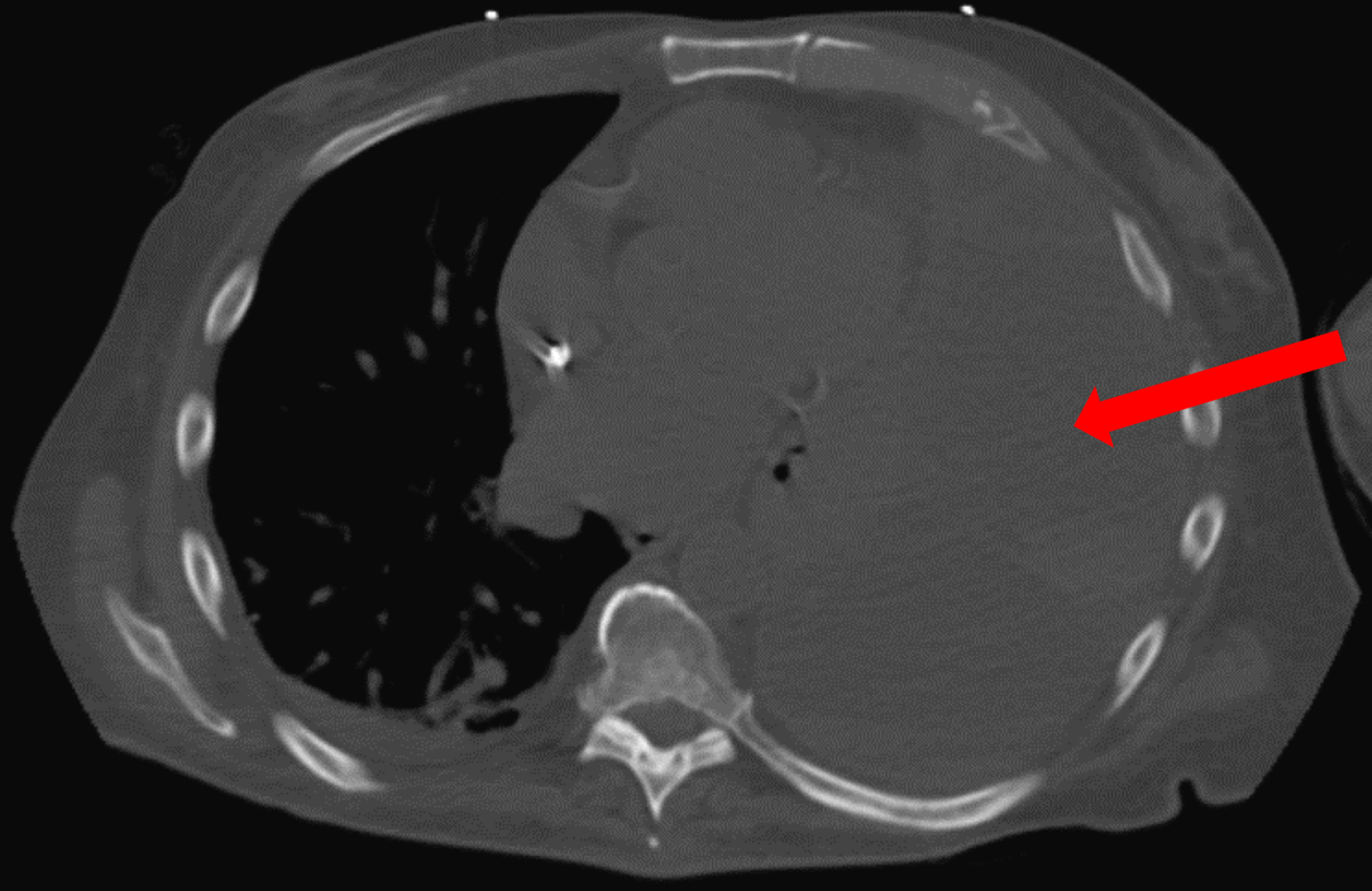
Comparative Interpretation Of Ct And Standard Radiography Of The Pleura from www.jbsr.be Jun 05, 2012 · thoracentesis in small or loculated pleural effusions, thereby increasing the yield and safety of the procedure. Due to the acute kidney injury, this exam was performed without contrast. Malignant pleural effusion and algorithm management. It is associated with significant morbidity and mortality. A pleural effusion describes an excess of fluid in the pleural cavity, usually resulting from an imbalance in the normal rate of pleural fluid production or absorption, or both. 34 the fluid may accumulate due to overproduction from diseased pleura, obstruction of lymphatic channels, or atelectasis of adjacent lung. The repeat ct again demonstrated a small loculated right pleural effusion with adjacent consolidation representative of an empyema. May 25, 2021 · the aetiology of the pleural effusion determines other signs and symptoms.
Dec 28, 2018 · zarogoulidis k, zarogoulidis p, darwiche k, et al.
Metintas m, ak g, dundar e, et al. May 25, 2021 · the aetiology of the pleural effusion determines other signs and symptoms. Dec 28, 2018 · zarogoulidis k, zarogoulidis p, darwiche k, et al. A computed tomography scan is very helpful if the lungs themselves are diseased. Jun 05, 2012 · thoracentesis in small or loculated pleural effusions, thereby increasing the yield and safety of the procedure. A pleural effusion describes an excess of fluid in the pleural cavity, usually resulting from an imbalance in the normal rate of pleural fluid production or absorption, or both. A loculated pleural effusion is not free flowing in the pleural space but rather. Malignant pleural effusion is the second most common cause of an exudative pleural effusion and the most common cause in patients over 60 years of age. Malignant pleural effusion and algorithm management. It is associated with significant morbidity and mortality. It has a higher yield than that of blind pleural biopsy in the diagnosis of malignancy. 34 the fluid may accumulate due to overproduction from diseased pleura, obstruction of lymphatic channels, or atelectasis of adjacent lung. Due to the acute kidney injury, this exam was performed without contrast.
A pleural effusion describes an excess of fluid in the pleural cavity, usually resulting from an imbalance in the normal rate of pleural fluid production or absorption, or both. A loculated pleural effusion is not free flowing in the pleural space but rather. It has a higher yield than that of blind pleural biopsy in the diagnosis of malignancy. Dec 28, 2018 · zarogoulidis k, zarogoulidis p, darwiche k, et al. However, it is not practical to recommend ultrasonography for all effusions.
Cureus Spontaneous Loculated And Massive Hemothorax An Uncommon Complication Of Warfarin Therapy from assets.cureus.com The repeat ct again demonstrated a small loculated right pleural effusion with adjacent consolidation representative of an empyema. Dec 28, 2018 · zarogoulidis k, zarogoulidis p, darwiche k, et al. Malignant pleural effusion and algorithm management. However, it is not practical to recommend ultrasonography for all effusions. Due to the acute kidney injury, this exam was performed without contrast. Metintas m, ak g, dundar e, et al. It is associated with significant morbidity and mortality. A pleural effusion describes an excess of fluid in the pleural cavity, usually resulting from an imbalance in the normal rate of pleural fluid production or absorption, or both.
A computed tomography scan is very helpful if the lungs themselves are diseased.
However, it is not practical to recommend ultrasonography for all effusions. May 25, 2021 · the aetiology of the pleural effusion determines other signs and symptoms. 34 the fluid may accumulate due to overproduction from diseased pleura, obstruction of lymphatic channels, or atelectasis of adjacent lung. It has a higher yield than that of blind pleural biopsy in the diagnosis of malignancy. Malignant pleural effusion and algorithm management. A pleural effusion describes an excess of fluid in the pleural cavity, usually resulting from an imbalance in the normal rate of pleural fluid production or absorption, or both. A loculated pleural effusion is not free flowing in the pleural space but rather. Dec 28, 2018 · zarogoulidis k, zarogoulidis p, darwiche k, et al. The repeat ct again demonstrated a small loculated right pleural effusion with adjacent consolidation representative of an empyema. A computed tomography scan is very helpful if the lungs themselves are diseased. Malignant pleural effusion is the second most common cause of an exudative pleural effusion and the most common cause in patients over 60 years of age. Jun 05, 2012 · thoracentesis in small or loculated pleural effusions, thereby increasing the yield and safety of the procedure. Metintas m, ak g, dundar e, et al.
Metintas m, ak g, dundar e, et al. May 25, 2021 · the aetiology of the pleural effusion determines other signs and symptoms. 34 the fluid may accumulate due to overproduction from diseased pleura, obstruction of lymphatic channels, or atelectasis of adjacent lung. A loculated pleural effusion is not free flowing in the pleural space but rather. Malignant pleural effusion and algorithm management.
Pleural Effusion from www.stritch.luc.edu Dec 28, 2018 · zarogoulidis k, zarogoulidis p, darwiche k, et al. A pleural effusion describes an excess of fluid in the pleural cavity, usually resulting from an imbalance in the normal rate of pleural fluid production or absorption, or both. A computed tomography scan is very helpful if the lungs themselves are diseased. The repeat ct again demonstrated a small loculated right pleural effusion with adjacent consolidation representative of an empyema. It is associated with significant morbidity and mortality. It has a higher yield than that of blind pleural biopsy in the diagnosis of malignancy. Malignant pleural effusion and algorithm management. 34 the fluid may accumulate due to overproduction from diseased pleura, obstruction of lymphatic channels, or atelectasis of adjacent lung.
If your doctor suspects a malignant pleural effusion, the next step is usually a thoracentesis , a procedure in which a needle is inserted through the chest wall into the pleural space to get a sample of the fluid.
Jun 05, 2012 · thoracentesis in small or loculated pleural effusions, thereby increasing the yield and safety of the procedure. Dec 28, 2018 · zarogoulidis k, zarogoulidis p, darwiche k, et al. 34 the fluid may accumulate due to overproduction from diseased pleura, obstruction of lymphatic channels, or atelectasis of adjacent lung. A pleural effusion describes an excess of fluid in the pleural cavity, usually resulting from an imbalance in the normal rate of pleural fluid production or absorption, or both. It has a higher yield than that of blind pleural biopsy in the diagnosis of malignancy. May 25, 2021 · the aetiology of the pleural effusion determines other signs and symptoms. The repeat ct again demonstrated a small loculated right pleural effusion with adjacent consolidation representative of an empyema. A computed tomography scan is very helpful if the lungs themselves are diseased. It is associated with significant morbidity and mortality. However, it is not practical to recommend ultrasonography for all effusions. If your doctor suspects a malignant pleural effusion, the next step is usually a thoracentesis , a procedure in which a needle is inserted through the chest wall into the pleural space to get a sample of the fluid. Metintas m, ak g, dundar e, et al. Due to the acute kidney injury, this exam was performed without contrast.